


How fertilisation takes place in humans
Fusion between the egg and the spermatozoon, step by step
Once the gametes are formed and in order to produce a new being, the egg and spermatozoon have to fuse. This process is called fertilization. Human fertilization is internal, that is, it takes place inside the woman's body, specifically in the fallopian tubes.
The egg or ovum is fertilised in the fallopian tube (1 day) and makes its way to the uterus (between 2 and 5 days). After five days, it reaches the uterine cavity where the embryo will nest (between 6 and 7 days). Fertilisation requires copulation, which consists of the introduction of the penis into the vagina and the subsequent ejaculation or expulsion of the semen.
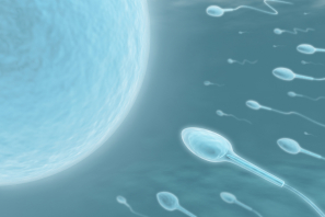
The semen deposited in the vagina goes through the uterus to reach the fallopian tubes. Within about two minutes of an ejaculation inside the vagina, the spermatozoa reach the end portion of the tubes. However, of the hundreds of thousands of spermatozoa, only a few will reach the egg and only one will be able to cross the egg's plasma membrane for it to be fertilised. All other spermatozoa are destroyed on the way. The reason for producing millions of spermatozoa is to ensure that at least one manages to reach the egg.
Once the spermatozoa have been deposited in the female reproductive system, as they ascend from the vagina, they suffer a capacitation phenomenon which consists of the partial loss of therostral portion of the spermatozoon head and an acrosome reaction, with small pores appearing at this level that release enzymes necessary for crossing the protective barriers of the oocyte.
At the time of ovulation, the ovary is partially covered by fimbriae tubae, that capture the released oocyte which adheres to the ovarian fossa, and carry it towards the uterus. The oocyte is in the so-called metaphase II (oocyte maturity stage) and is surrounded by the corona radiata and the zona pellucida.
Fertilization, step by step
The fertilization process requires the following steps:
-
Penetration of the corona radiata
Of the 200 or 300 million spermatozoa deposited in the vagina, only between 300 and 500 reach this stage of the fertilisation process. Once inside the corona radiata, it would appear that the hyaluronidase (capable of hydrolysing hyaluronic acid, a mucopolysaccharide which is abundant in the zona pellucida and in the cementing substance of the follicle cells) causes the dispersion of cells in the corona. However, nowadays, it is believed that these cells are dispersed by the combined action of sperm enzymes and tube mucous. -
Penetration of the zona pellucida
This second barrier is crossed with the help of enzymes, called sperm lysins, released by the acrosome. The release of these sperm lysins is associated with a series of structural changes in the spermatozoon, mainly affecting the acrosome. This is known as an acrosome reaction. Together, these changes form what is known as the sperm activation process. This process is triggered by substances diffused from the egg such as those released from the acrosome granule, which could correspond to sperm lysins. From the remaining parts of the acrosome, what is known as acrosome filament, which develops into the activated spermatozoa, begins to grow.
By means of flagellar movements, the spermatozoon pushes the acrosome filament until it makes contact contacts with the egg’s cell membrane. Many spermatozoa do not undergo the acrosome reaction until they have bound to glycoprotein receptors in the zona pellucida. After penetrating one of these receptors, the permeability of the membrane is modified by a process called zone reaction.
-
Penetration of the oocyte’s plasma membrane
The union of the first spermatozoon with the plasma membrane triggers three events: as soon as the sperm comes into contact with the oocyte membrane, the plasma membrane of that spermatozoon fuses with the oocyte’s plasma membrane in the fertilization cone, and the head, the mid-piece and tail of the sperm enter the oocyte cytoplasm, leaving the plasma membrane behind on the surface of the oocyte. Once inside, the oocyte ends its second meiosis, releasing a second polar body and the chromosomes are placed in a vesicular nucleus called the female pronucleus. At the same time, the oocyte contracts and a space appears between the oocyte and the zona pellucida, This is called the perivitelline space. The spermatozoon advances until it is next to the female pronucleus and its nucleus swells forming the male pronucleus. Then, the tail detaches from the head and degenerates.- Fertilization cone formation or egg cytoplasmic protrusion.
- Ion changes (calcium, sodium and hydrogen) and cytosol cause instantaneous and temporary depolarisation of the membrane.
The cortical granules release their content into the space around them. This cortical reaction alters the glycoprotein receptors in the zona pellucida, blocking the adhesion of more spermatozoa to the egg.
Before the fusion of the two pronuclei (haploid and 1n DNA, i.e. with 23 chromosomes), they must each duplicate their DNA. Otherwise, each cell of the zygote in the bicellular state would have half as much DNA as normal.
After DNA synthesis, the chromosomes are placed in the spindle and the 23 paternal and 23 maternal chromosomes are split lengthwise at the centromere (as in normal mitotic division). The resulting halves are then randomly segregated and move to the opposite poles, giving each cell the normal number of chromosomes and DNA (2n).
The cell unites in its central zone and the cytoplasm is divided into two parts. The now-fertilizated egg called a zygote is a single-cell containing 46 chromosomes, as it has the 23 chromosomes of the oocyte plus the 23 of the spermatozoon. It will begin a "return" journey until it implants in the uterus.